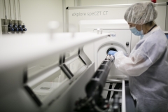
At its Molecular and Functional Imaging Facility, biomaGUNE, the Centre for Cooperative Research devoted to state-of-the-art research into biomaterials, has resources that the scientific community can avail itself of. This facility occupying an area of 900 m2 is an integrated bioimaging resource run by expert professionals and offers state-of-the-art preclinical imaging equipment, such as Positron Emission Tomography (PET), Single Photon Emission Computerized Tomography (SPECT), Computerized Tomography (CT), Magnetic Resonance Imaging (MRI) and optical imaging. It also has a radiochemical laboratory with a biomedical cyclotron, advanced microscopy equipment and an Animal Facility Unit for small rodents accredited by the Association for the Assessment and Accreditation of Laboratory Animal Care (AAALAC).
CIC biomaGUNE’s Molecular and Functional Imaging Facility, recognised by the Spanish Ministry of Science, Innovation and Universities as an Outstanding Scientific and Technological Infrastructure (ICTS) in biomedical imaging, has been designed to develop innovative research projects in the field of preclinical molecular and functional imaging and in the field of nanomedicine. It is open to collaborating with research groups as well as with national or international companies by offering the knowledge, infrastructure and experience it has at its disposal for the conducting of longitudinal, multimodal preclinical projects.
Jordi Llop, lead researcher at CIC biomaGUNE's radiochemistry and nuclear imaging laboratory, stresses that “all imaging techniques, a cyclotron and a radiochemistry laboratory have been brought together under one roof —under one management—; all kinds of contrast agents and tracers for in situ research can be conducted here, and that gives us the capacity to tackle studies in addition to huge flexibility and speed”. The facility also has an 11.7-tesla MRI scanner “the most powerful in its class and the only one in the whole of the Iberian Peninsula”.
Longitudinal, multimodal studies for a whole range of diseases
Research centres, universities, hospitals and companies that need “to conduct non-invasive studies, in vivo studies in which the evolution of small rodents is monitored over time by combining various imaging techniques, can make use of various possibilities at the facility. It should be highlighted that we are not geared towards a single disease; instead, we tackle and are experts in projects relating to neurodegenerative, cerebrovascular, oncological, cardiovascular, pulmonary diseases, metabolic disorders, toxicology, etc. Besides having a catalogue of about 30 to 40 radiotracers, there is an option to produce others tailored to each project, because every disease is looked at differently,” pointed out Dr Llop. The facility also has experts in the development of nano-radiotracers and allows nanoparticle systems with theragnostic applications (i.e., ones for both therapeutic and diagnostic purposes) to be used.
There are many applications, but in general terms Jordi Llop sums up the activity of the facility thus: “In the process to develop drugs it is necessary to carry out target testing (to test whether the drug reaches the intended location), mechanism testing (to show that the drug activates or hinders reactions as expected), and effectiveness testing (to show that the drug has the expected healing effect). Likewise, it is possible to study how a specific disease affects specific mechanisms of the body. In vivo imaging techniques offer the chance to conduct longitudinal studies, and that facilitates the transfer of these studies to the clinical environment, in other words, it will be easier to move them on to the clinical phase in humans.”
Various studies are being developed right now. In one of them the “therapeutic efficacy of a drug for Alzheimer’s disease is being monitored. We have another study in collaboration on autism; we are also working on the study of a peptide that can be selectively built up in specific tumours and could have an application in radiotherapy; and we have just established a partnership to monitor whether a kind of nanobody with the potential for curing Alzheimer’s can cross the blood-brain barrier and reach the brain”.
In addition, applications on an anatomical, functional and metabolic level of imaging techniques are being developed in the study of the central nervous system at the Centre’s Magnetic Resonance Imagin Lab; this is in line with the quest for new diagnostic techniques in diseases relating to this system; nanomedicines for the advanced treatment of cerebrovascular, neurodegenerative diseases, etc. are also being developed. And in the Molecular and Functional Biomarker Laboratory they are specialised in cardiovascular and pulmonary imaging, and, among other things, are developing imaging and molecular biomarkers for the early diagnosis of pulmonary hypertension, and these biomarkers are being used to monitor the efficiency of new therapies.
External researchers can access these outstanding scientific and technological resources through a competitive call that has been opened (through the Distributed Biomedical Imaging Network, ReDiB), which offers much more affordable prices than if it was just a direct service. “We believe that it is important to make our facility known because potential users are unaware of the resources they can avail themselves of, or the type of projects that can be tackled here,” concluded Dr Llop.

